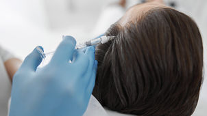
What are Skin Biopsies?
Skin Biopsies are medical procedures that involve removing a small sample of tissue from the body for examination under a microscope. This diagnostic tool helps identify skin conditions, confirm cancer diagnoses, and guide treatment plans by providing detailed information about cellular structures and abnormalities.

Our Approach to Skin Biopsies
Stage 1
Thorough Evaluation and Preparation
We begin by assessing the patient’s medical history and current symptoms to determine the need for a biopsy. Our dermatologist will explain the procedure, addressing any concerns and ensuring patients are informed and comfortable with the biopsy process.
Stage 2
Tissue Sampling
We perform the biopsy, selecting the most appropriate method, such as punch, shave, or excisional biopsy. Local anesthesia ensures minimal discomfort while obtaining a tissue sample for comprehensive pathological analysis.
Stage 3
Detailed Analysis and Follow-Up
The tissue sample is analyzed by a specialized pathologist to diagnose potential conditions. We discuss the results with the patient, providing a clear explanation and recommending a personalized treatment plan, ensuring ongoing support and optimal care throughout the treatment journey.

Benefits of Undergoing Skin Biopsies
Accurate Diagnosis
Skin biopsies provide precise diagnosis, identifying skin conditions or confirming cancer, guiding effective treatment plans for optimal patient outcomes.
Early Detection
Early detection through skin biopsies enables timely intervention, improving prognosis and reducing potential complications associated with untreated skin conditions.
Skin Health Insight
Skin biopsies offer detailed insights, allowing dermatologists to tailor personalized treatment strategies, ensuring targeted care and improved long-term skin health.
When Skin Biopsies Become Necessary
Understanding various skin conditions can help individuals identify when to seek medical advice.
-
Persistent, unusual lesions require examination
-
Altered size, shape, or colour evaluated
-
Persistent, unresponsive rashes investigated
-
Confirm diagnosis through tissue analysis
-
What types of skin cancer can be treated with Mohs Micrographic Surgery?Mohs Micrographic Surgery is primarily used for basal cell carcinoma and squamous cell carcinoma. It is also effective for other skin cancers in areas where preserving healthy tissue is crucial, such as the face.
-
How long does the Mohs Micrographic Surgery procedure take?The procedure can take several hours, as each layer of tissue is removed and analyzed on-site until clear margins are achieved. The exact duration depends on the cancer's size, location, and depth.
-
What should I expect during the recovery period?Recovery is usually quick, but it can vary based on the cancer's size and location. Patients may experience some discomfort, swelling, or bruising. Following your surgeon's post-operative care instructions is essential for optimal healing.
-
Are there any risks or complications associated with Mohs Micrographic Surgery?As with any surgery, there are potential risks, such as bleeding, infection, scarring and recurrence. Your Mohs Micrographic surgeon will discuss these with you.
-
How should I prepare for Mohs Micrographic SurgeryPatients should discuss any medications they are taking with their surgeon and follow specific pre-operative instructions provided. It is recommended to arrange transportation home after the procedure due to potential temporary discomfort or numbness.
-
What are the benefits of Mohs Micrographic Surgery compared to other skin cancer treatments?Mohs Micrographic Surgery offers a high cure rate and minimizes the removal of healthy tissue, which leads to better cosmetic outcomes. It is ideal for cancers with a high risk of recurrence or those located in cosmetically sensitive areas.
Relevant Articles On Skin Biopsies


